Pre-therapeutic evaluation
After the diagnosis of a lymphoma, various complementary investigations may be conducted to study the prognostic factors and guide the choice of treatment
Examples of complementary investigations that may be required:
- Biological blood tests
- Medical imaging investigations
- Medullary biopsy or bone biopsy
- Lumbar puncture
- Gastric fibroscopy
- Heart examinations
Biological blood tests
Various biological blood tests provide information about the disease and its extent, as well as the general state of health of the patient.
Examples of factors that are studied:
- The concentration of lactate dehydrogenase (LDH) in the blood is the most frequently used. It indicates the size of the tumour and its aggressiveness.
- The concentration of beta-2 microglobulin is similar, but less frequently used.
- The concentration of albumin in the blood reflects the general state of health, and in particular the state of nutrition of the patient.
- The increase of the sedimentation rate or any other element that indicates inflammation is a worrying prognostic element in Hodgkin lymphomas.
- The decrease of haemoglobin concentrations, which indicates anaemia, may be a prognostic factor.
Medical imaging investigations
Medical imaging investigations are performed in order to evaluate the extent of the lymphoma. These are painless investigations. The choice of the medical imaging technique or techniques to be used depends on the type of lymphoma suspected.
CT scan or tomodensitometry
This is a radiological examination based on the use of X-rays. Several images are taken from various angles, which make it possible to form a three-dimensional image on the computer. These scans are usually performed on the whole of the thorax and the abdomen in order to determine which lymph glands are affected and their size, as well as other potentially affected organs.
A scan of the whole thorax and abdomen is always performed in the initial evaluation of a lymphoma. It will be performed anew during the treatment and at the end of the treatment, in order to determined the response of the disease to the treatment. It will then often be proposed as a monitoring examination at regular intervals.
A scan of the thorax that shows abnormal mediastinal nodes.

PET-Scan (Positron Emission Tomography)
This is an examination that makes it possible to detect cancer cells and track their evolution. To do so, a weakly radioactive product, most frequently 18F-FDG, is injected intravenously. This product primarily fixes onto cancer cells. Its spread across the organism thus makes it possible to detect these cells. The PET scan can be performed to detect cancer tissue or to verify its absence during the diagnosis of the disease, during the treatment and in the context of after-treatment monitoring.
PET scans are becoming more and more important in the evaluation or certain lymphomas, especially aggressive lymphomas. It is particularly useful to evaluate the response of the disease to a treatment.
Medullary biopsy or bone biopsy
A medullary biopsy, or bone biopsy, is a bone marrow sample that is taken in order to examine it. The bone marrow, which is the tissue located within the bones, contains the immature blood cells that will then develop into white blood cells, red blood cells and platelets. The bone marrow is often affected by lymphomas, which is why a medullary biopsy is usually necessary in the initial evaluation.
The sample is usually taken under local anaesthetic. It does not require hospitalisation, and takes approximately 15 minutes. A short general anaesthesia with a gas (nitrous oxide) is now often proposed to the patient in addition to the local anaesthesia for more comfort. After the local anaesthesia is applied to the pelvic bone, the sample is taken. This is performed in two stages: aspiration of a drop of bone marrow, then an actual bone biopsy with a biopsy needle.
This sample is usually well tolerated and causes little pain. When the effect of the anaesthesia wears off, the sample point may become tender, at which point painkillers may be prescribed by the doctor.
The sample analysis usually takes 8 to 10 days. The results are then sent directly to the doctor who prescribed the examination.
Lumbar puncture
In certain lymphoma cases, a lumbar puncture may be necessary. This examination requires taking a few millilitres of the liquid that surrounds the spinal cord and the brain (cerebrospinal fluid or CSF).
This examination may be necessary in the initial disease evaluation in order to search for cancer cells. It can also be used in certain cases during the treatment, if there is the risk of a relapse affecting the spinal cord. A lumbar puncture makes it possible to inject chemotherapy at that point.
This examination is performed under local anaesthetic. A long, very thin needle is introduced into the base of the spine by the doctor in order to take a sample of the liquid. This liquid will then be examined under the microscope at the laboratory. Then, if necessary, the doctor will inject a dose of chemotherapy at the same spot.
This examination does not require hospitalisation and is usually painless. The patient is required to remain lying flat for one to three hours in order to avoid headaches. Indeed, certain patients may suffer from headaches in the hours or days after the puncture, which may last for several days and require strong painkillers and bed-rest.
Gastric fibroscopy
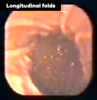
A gastric fibroscopy is performed by introducing a thin tube into the digestive tract (oesophagus then stomach), making it possible to see into it. This examination is proposed if there is a doubt about the presence of cancerous cells in the stomach.
It usually does not require anaesthesia. It is not painful, but it can be rather uncomfortable. It is necessary, of course, to have not eaten or drunk for several hours before the examination. It is important to try and be well relaxed before and during the examination.
Heart examinations
Certain medicines used in lymphoma treatments are toxic for the cardiac muscle. It is sometimes preferable to avoid them for patients who already suffer from a heart disorder. An examination of the cardiac function is therefore necessary to ensure they can be used.
The examination of the cardiac function makes it possible to calculate the “systolic ejection fraction” (SEF), which measures the contraction capacity of the left ventricle. This measure can be taken in two ways: through cardiac ultrasound or through isotopic measurement. The first is a simple ultrasound. The second requires the injection of a radioactive marker and the examination of its passage into the heart with a special camera (scintigraphy).